Spatial Transcriptomics
Spatial transcriptomics combines high-plex quantification with the spatial resolution of immunohistochemistry. This technology classifies tissue based on total mRNA within the morphological context of FFPE or frozen tissues. Spatial expression mapping is a powerful tool for examining cellular interactions, tissue and tumor heterogeneity, pathogenicity, and response to therapy.
GENEWIZ offers spatial profiling utilizing the 10X Genomics® Visium HD® platform. This novel technology performs simultaneous, in situ spatial analysis from a single FFPE or frozen tissue section. You can select from a host of discovery-focused panels with the potential for customization. Together with our epigenetic and single-cell sequencing solutions, you have access to the most cutting-edge technologies available to study gene expression from every angle and every level.

Why is spatial profiling important?
Digital spatial profiling (DSP) is important for analyzing cellular heterogeneity and intracellular interactions, especially in the context of drug therapy response. Important advantages of DSP:
- Samples can be stored and probed again
- Multiple regions of interest (ROI) can be explored from a single FFPE slide
- DSP offers the benefits of both IHC/FISH and high-throughput expression analysis
10x Genomics Visium HD Spatial Transcriptomics
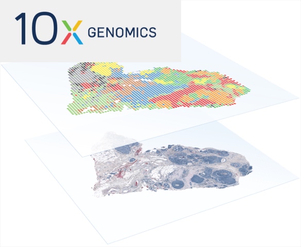
High-resolution full mapping of the transcriptome across tissue sections while preserving spatial context
- Minimal workflow optimization needed
- No need to select regions of interest (ROI)
- Integrates histological imaging with gene expression data to provide tissue architecture and cellular organization insights
- Compatible with FFPE and fresh-frozen samples, with the ability to process archival H&E-stained pathology slides
- Ideal for studying heterogeneity, developmental biology, disease progression, and cellular interactions within single-cell resolution and at scale
Applications of Spatial Transcriptomic Technology
-
Oncology
- Profile tumor heterogeneity, microenvironment, and cancer stem cells
- Identify tumor-infiltrating lymphocytes (TILs)
-
Immunology
- In situ immune repertoire characterization
- Immune response profiling of normal and diseased states
-
Neuroscience
- Contextually profile neural and stem cells
- Characterization of neurodegenerative disease pathology
-
Pathology
- High-plex analysis of expression in normal and diseased tissues
- Biomarker discovery and characterization
- Monitor therapy response
-
Embryo Development
- Characterize spatial gene expression during embryogenesis
- Associate gene expression with pluripotency and cellular differentiation
Technical Resources
-
Tech Note | High-Resolution Spatial Profiling and Biomarker Discovery by Visium HD
In this tech note we explore how the spatial transcriptomics platform, Visium HD®, bridges the gap between histology and gene expression profiling, offering precise biomarker discovery and cellular mapping within complex tissues to support research across oncology, immunology, and developmental biology.
-
eBook │ A Guide to Single-Cell Sequencing
Whether you’re new to single-cell sequencing or looking to improve your data quality, this eBook provides a comprehensive overview of single-cell sequencing, focusing on how this technology works, optimized approaches to enhance results and use-case applications to help you get the most out of your research.
-

Webinar | Advancing Transcriptomics: Gene Expression Screening, Single-Cell RNA-Seq, and Beyond
With this two-part webinar series, go beyond traditional transcriptomics and learn about the various NGS approaches available for gene expression analysis. In part 1, we take an in-depth look at various gene expression approaches. In part 2, we explore the data generated from these approaches and how they can complement each other and confirm findings.
-
Tech Note | Enhanced Gene Detection with GEM-X Single-Cell Sequencing
This tech note highlights how 10x Genomics’ GEM-X chemistry enhances single-cell RNA sequencing by improving cell recovery, gene detection, and immune profiling, while reducing costs per cell.
NGS Platforms
For information on our NGS platforms as well as recommended configurations of your projects, please visit the NGS Platforms page. GENEWIZ does not guarantee data output or quality for sequencing-only projects.